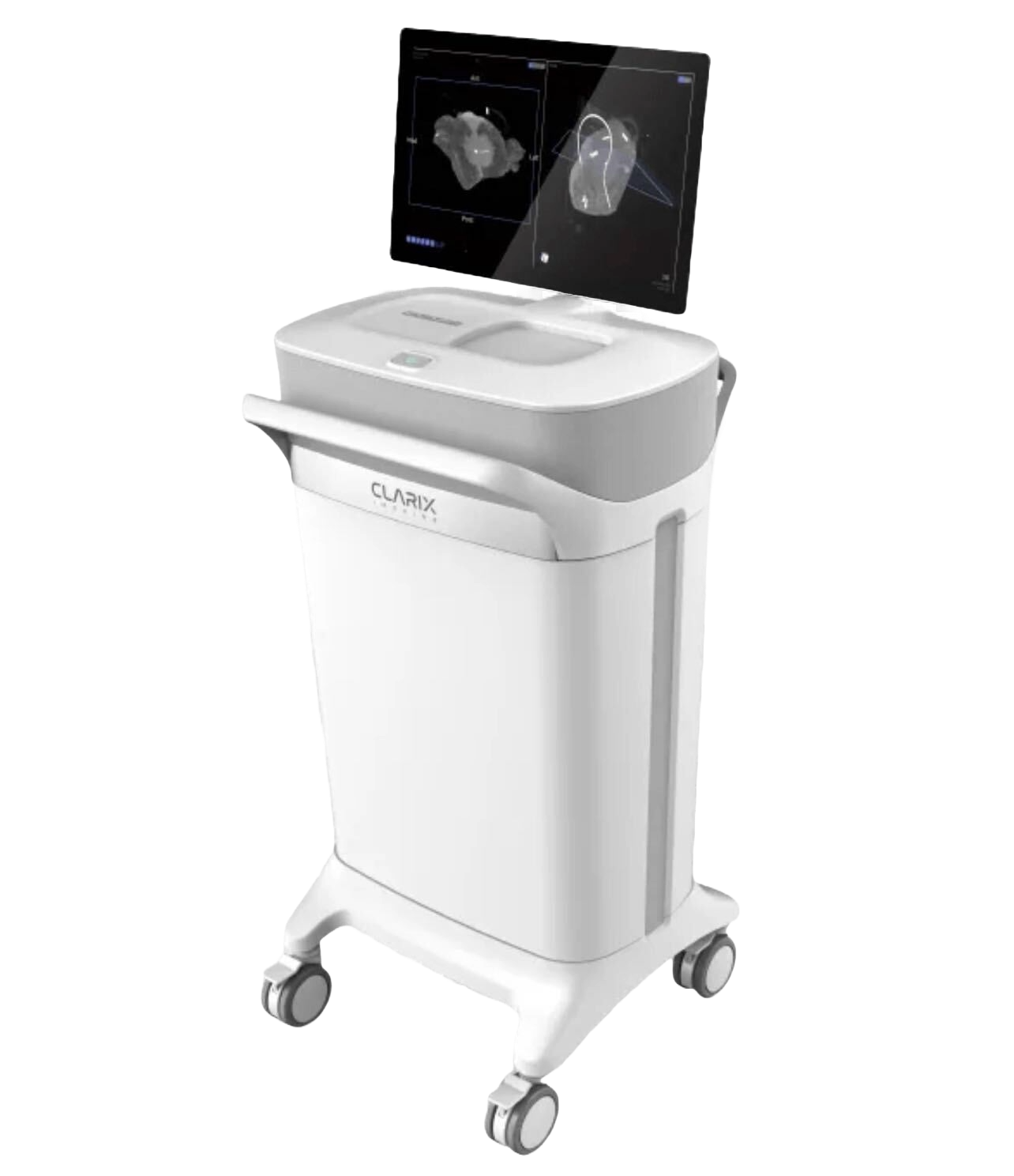
True 3D specimen imaging for precision breast conserving surgery
Transform surgical outcomes with Clarix Imaging's advanced 3D intraoperative imaging technology. Real-time visualization that empowers surgeons to make confident, precise decisions.
⚡ We will take you soaring through the specimen, showing the scan in 3D
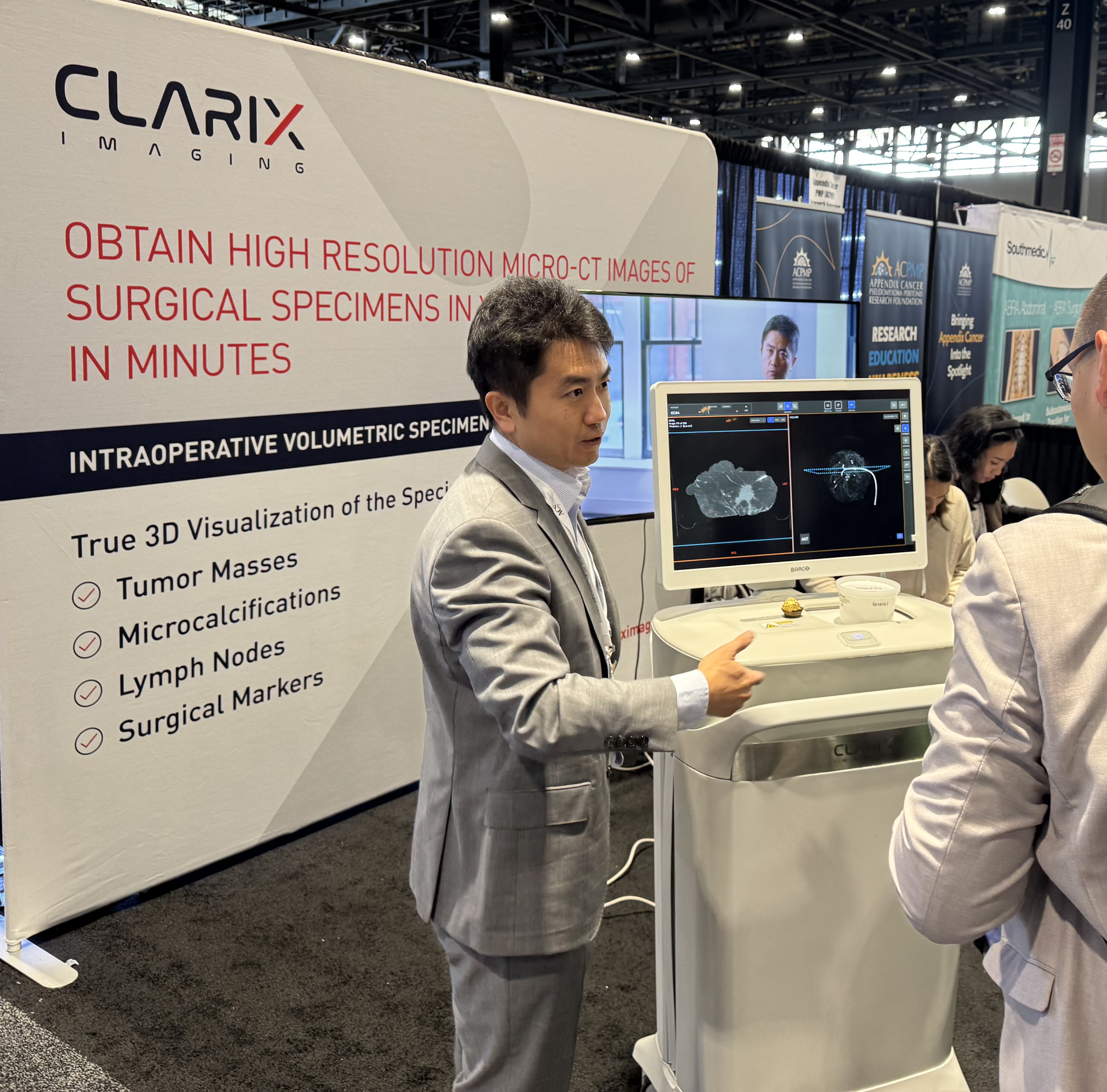
Sophisticated technology, simplified workflow
Clarix Imaging's cutting-edge computational imaging technology is seamlessly integrated into your current surgical workflow, enabling real-time insights when they matter the most.
True 3D clarity
Volumetric image reconstruction provides the highest 3D resolution and contrast, revealing tiny details for unprecedented visualization clarity and precise 3D orientation tracking.
Intuitive 3D navigation
Interactive visualization of tomographic image slices and volumetric rendering – exactly the same way you view CT or MR images requiring little training.
Uninterrupted workflow
Immediate availability of 2D scout image for surgical marker confirmation. <3 min lightning scan and image processing saves valuable OR time.
Outcomes that matter to surgeons and patients
Reduce Positive margins
Real-time visual examination of every margin helps achieve clear margins in the first procedure, reducing positive margins and re-excisions. (Kaweah Health study)
Enhanced confidence
Make confident decisions for optimized breast conservation, with less amount of tissue removed while minimizing positive margins.
Streamlined workflow
No need to change your existing OR workflow.
Experience the future of intraoperative imaging
Join leading breast cancer surgeons who trust Clarix Imaging to deliver optimized breast conserving surgery in every procedure.